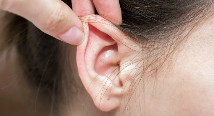
The anatomical projection and symmetry of the ears play a major role in framing the facial profile. When ears project more than 2 centimeters from the side of the head (a condition clinically recognized as prominent ears), it often creates severe psychological distress, teasing in childhood, and chronic self-consciousness in adulthood. Many individuals adapt their lifestyles by avoiding short haircuts, refusing to wear their hair tied back, or feeling insecure during social interactions. While temporary splints work exclusively on soft newborn cartilage, adult ears feature fully developed, elastic cartilage that cannot be reshaped without surgical repositioning. In modern facial plastic and reconstructive surgery, prominent ear correction (otoplasty) provides a permanent, harmonious solution for setting ears back.
At Stamboul Clinic, we coordinate world-class ear plastic and reconstructive procedures within accredited surgical hospital settings. Whether you are researching an all-inclusive prominent ear correction Turkey itinerary, seeking clarity on the possibilities of prominent ear correction without surgery, examining real prominent ear correction before after milestones or understanding prominent ear correction recovery, this guide delivers deep clinical data.
Table of Content
- What Is Prominent Ear Correction?
- Surgical vs. Non-Surgical Options: Prominent Ear Correction Without Surgery
- Incisions and Scar Placement: Are Otoplasty Scars Visible?
- Timeline and Headband Care: Prominent Ear Correction Recovery
- Prominent Ear Correction Before After Results
- Frequently Asked Questions (FAQ)
- How do I sleep safely during the first month after prominent ear correction surgery?
- Will prominent ear correction surgery affect my hearing or inner ear health?
- Can prominent ears pop back out or relapse after otoplasty surgery?
- At what age can children undergo prominent ear correction surgery?
- When can I safely wash my hair, wear glasses, and return to sports after surgery?
What Is Prominent Ear Correction?
To establish a clear medical foundation, let us examine what happens during prominent ear correction surgery from a structural perspective. Clinically known as otoplasty, it is a delicate cosmetic and reconstructive procedure performed under local anesthesia with sedation or general anesthesia.
THE 2 PRIMARY ANATOMICAL CAUSES OF EAR PROTRUSION:
- Underdeveloped Antihelical Fold ➡️ The outer upper rim of the ear fails to fold back, causing the top to stick out.
- Hypertrophic Conchal Bowl ➡️ Excessive, deep cartilage in the ear cup physically pushes the entire ear forward.
In over 80% of clinical cases, prominent ears result from a combination of an unformed antihelical fold and an overgrown conchal cartilage bowl. During surgical correction, the surgeon addresses both structural deficits: scoring or folding the cartilage to create a smooth antihelix and setting the deep concha back toward the mastoid bone behind the ear.
Surgical vs. Non-Surgical Options: Prominent Ear Correction Without Surgery
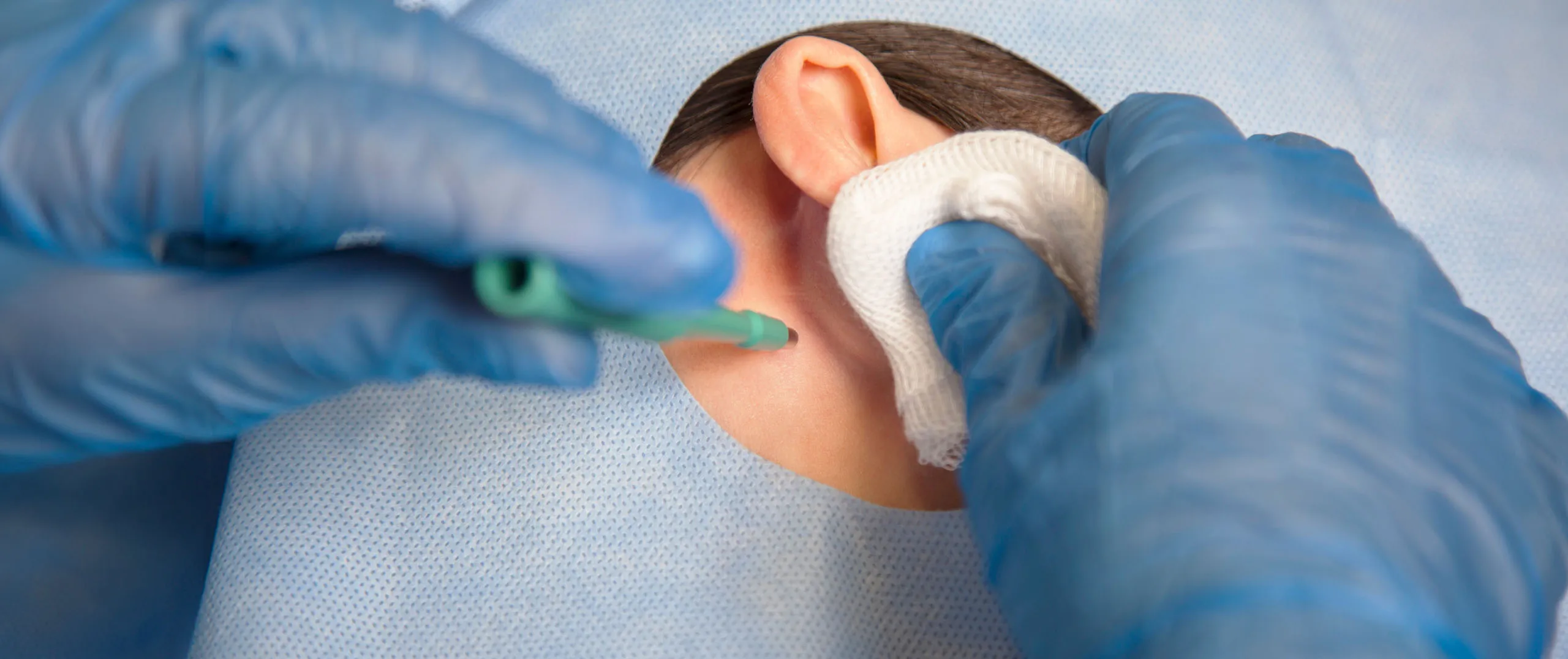
Understanding the physical limitations of non-surgical techniques helps patients choose the appropriate corrective path.
TREATMENT MODALITIES BY AGE AND CARTILAGE MATURITY:
- Neonatal Ear Molding (Non-Surgical): Effective ONLY during the first 6 weeks of life while maternal estrogen keeps cartilage soft.
- Incisionless Thread Otoplasty: Places hidden internal sutures without skin cuts; high relapse rate if cartilage springs back.
- Traditional Cartilage Otoplasty: Gold standard surgical procedure; permanent structural reshaping with zero relapse.
The Reality of Prominent Ear Correction Without Surgery
Non-surgical ear molding devices (such as silicone splints and ear tapes) are clinically effective only within the first 6 to 8 weeks after birth. During this brief neonatal window, maternal estrogens circulating in the infant's bloodstream keep the cartilage malleable. Once maternal estrogen drops, the cartilage hardens permanently. In older children and adults, external tapes or glues only offer a cosmetic hold for a few hours and frequently trigger skin dermatitis without altering cartilage structure.
Surgical Cartilage Reshaping (Mustarde & Furnas Sutures)
Permanent correction requires a surgical approach. The surgeon makes a discrete incision on the posterior surface (the back) of the ear. Using non-absorbable Mustarde sutures, the cartilage is gently folded to build a defined antihelix. Permanent Furnas sutures are then placed to anchor the conchal cup directly to the mastoid fascia behind the ear, reducing the ear-to-head projection angle to a natural 15 to 20 degrees.
Incisions and Scar Placement: Are Otoplasty Scars Visible?
Managing patient expectations regarding surgical scarring is a key component of pre-operative planning.
THE POST-OPERATIVE SCAR SEQUENCE:
- [Incision Hidden in Retroauricular Sulcus] ➡️ [Zero Skin Cuts on Front of Ear] ➡️ [Dissolvable Internal Sutures] ➡️ [Scar Matures into Faint Line]
Because the surgical incision is placed exclusively within the natural crease behind the ear (the retroauricular sulcus), there are zero visible scars on the front or sides of the ears. Once fully healed over 3 to 6 months, the incision fades into a thin, pale white line hidden within the shadow of the ear crease.
Timeline and Headband Care: Prominent Ear Correction Recovery
Navigating your prominent ear correction recovery with clarity ensures stable cartilage healing and prevents accidental trauma.
THE POST-OPERATIVE RECOVERY TIMELINE:
- Days 1 to 3: Acute Bandage Phase ➡️ Bulky protective ear dressing worn; mild throbbing managed with pain medication.
- Day 7: Dressing Removal ➡️ Bulky wrap replaced with a soft, elastic sports headband; return to light desk work.
- Weeks 2 to 4: Night-Time Headband ➡️ Headband worn exclusively during sleep to prevent ears from folding forward in bed.
- Months 2 to 3: Cartilage Integration ➡️ Full cartilage tensile strength restored; contact sports safely resumed.
Patients must wear a soft, elastic tennis headband 24/7 during week 1 to keep the newly positioned cartilage splinted against swelling. For the subsequent 3 weeks, the headband is worn solely during sleep to prevent the ears from bending or pulling forward against bed pillows.
Prominent Ear Correction Before After Results
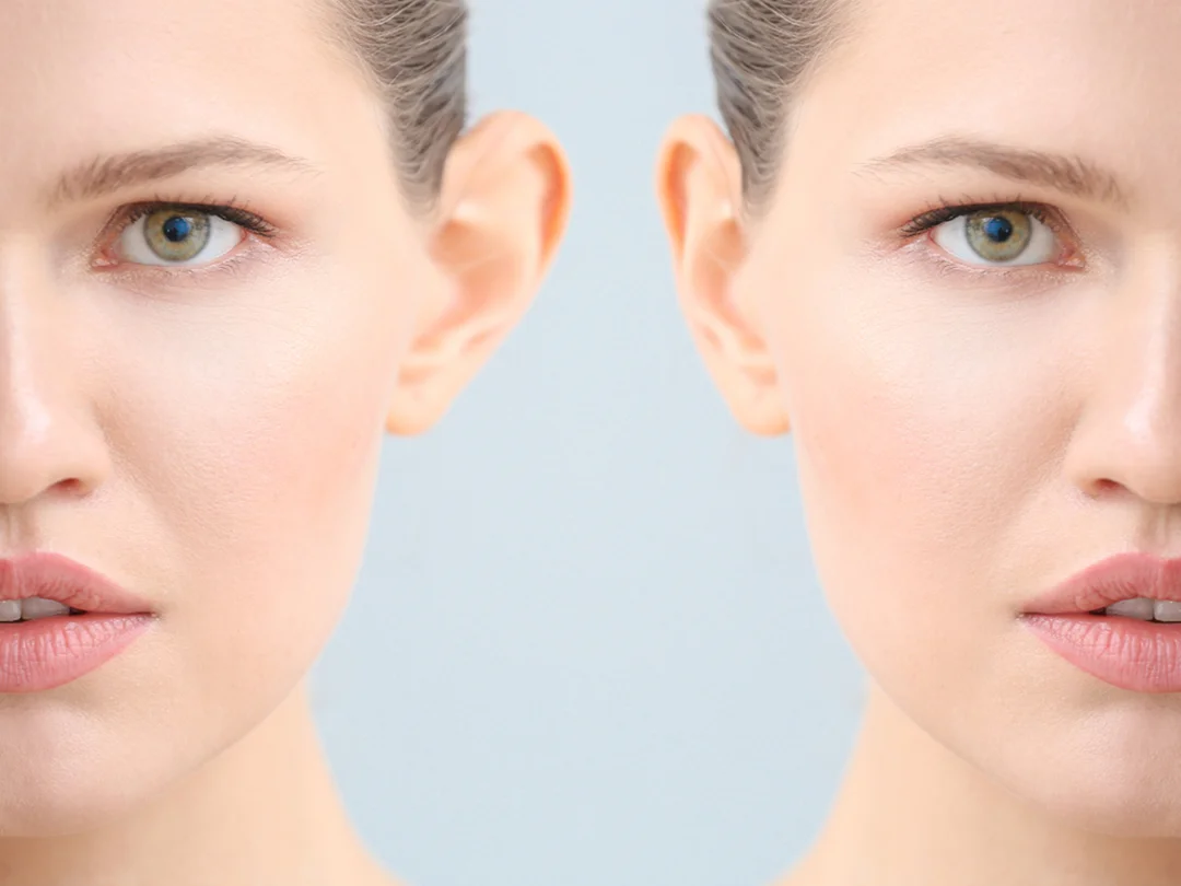
Evaluating genuine prominent ear correction before after patient profiles illustrates how natural symmetry and anatomical repositioning restore facial balance:
Modern otoplasty techniques avoid the artificial, "pinned-back" look created by outdated cartilage-cutting methods. By combining gentle cartilage scoring with tension-free suture suspension, surgeons preserve the natural contours of the ear rim without sharp cartilage ridges.
Frequently Asked Questions (FAQ)
How do I sleep safely during the first month after prominent ear correction surgery?
You must sleep strictly on your back with your head elevated at a 30-to-45-degree angle on 2 soft pillows for the first 3 to 4 weeks. Never sleep on your side, as direct pressure on the ears can displace the internal cartilage sutures. Always wear your protective elastic headband during sleep to prevent the ears from catching and folding forward on the pillow.
Will prominent ear correction surgery affect my hearing or inner ear health?
No. Prominent ear correction operates entirely on the external auricular cartilage and skin. It does not touch the ear canal, middle ear bones, or inner ear acoustic nerve mechanisms. Your hearing function remains unaffected.
Can prominent ears pop back out or relapse after otoplasty surgery?
When performed using permanent, non-absorbable internal cartilage suspension sutures (Mustarde and Furnas techniques), the long-term relapse rate is extremely low (under 3%). Following your post-operative headband instructions for the full 4 weeks ensures that biological scar tissue matures securely around the sutures.
At what age can children undergo prominent ear correction surgery?
Children can safely undergo prominent ear correction starting at age 5 to 6. By this age, the ear cartilage has reached approximately 85% to 90% of its adult size and is firm enough to hold internal sutures, allowing parents to correct protruding ears before the child enters primary school to prevent teasing.
When can I safely wash my hair, wear glasses, and return to sports after surgery?
You can gently wash your hair 3 to 4 days after surgery once your initial post-op checkup is complete, ensuring the area behind the ears is dried immediately with a cool hairdryer setting. Avoid resting heavy eyeglass frames directly on the incision crease for 2 weeks (tape frames slightly above the ears if needed). Strenuous workouts and contact sports must be avoided for 4 to 6 weeks.
Über den Autor