
The lateral canthus, the outer anatomical corner where the upper and lower eyelids meet, defines the horizontal palpebral aperture, eye slant, and periorbital vitality. In an aesthetically balanced, youthful orbit, the lateral canthus sits approximately two to four millimeters higher than the medial (inner) canthus, creating a natural upward tilt. However, chronological aging, congenital laxity, facial nerve weakness, or complications from previous over-resected lower blepharoplasty procedures can weaken the lateral canthal tendon.
When this support structure stretches, the lower eyelid drops downward, exposing the white sclera below the iris (scleral show) and producing a rounded, fatigued, or sad "hound-dog" expression. Severe laxity can even cause the eyelid to roll outward (ectropion), leading to chronic dry eye and tearing. While temporary thread lifts dissolve within months, lasting periorbital reconstruction requires surgical tendon anchoring. In modern oculoplastic and facial plastic surgery, canthoplasty provides the definitive surgical standard to reconstruct lower lid suspension and sculpt a refined almond eye shape.
Table of Content
- What Is Canthoplasty?
- Canthoplasty vs. Canthopexy: The Structural Difference
- Aesthetic vs. Functional Indications: Almond Eye Surgery and Ectropion Repair
- Incision Design and Scar Concealment: Are Canthoplasty Scars Visible?
- Recovery Timeline: Navigating Healing Milestones and Chemosis
- Visual Proof: Canthoplasty Before and After Transformations
- Cost Determinants: Understanding Canthoplasty Pricing Variables
- Frequently Asked Questions (FAQ)
- Will my eyes look artificially slanted or pulled right after canthoplasty?
- What is the primary difference between a canthoplasty and a blepharoplasty?
- When can I safely wear contact lenses after canthoplasty surgery?
- Can canthoplasty correct a round eye shape caused by a previous bad surgery?
- Why do canthoplasty prices vary between different patients?
What Is Canthoplasty?
To establish a clear medical foundation, let us examine the primary question: what is canthoplasty from an oculoplastic and ophthalmic perspective? Clinically defined as a surgical reconstruction of the lateral canthal angle, canthoplasty surgery is an outpatient micro-procedure performed under local anesthesia with light twilight sedation or general anesthesia lasting roughly one to two hours.
THE LATERAL CANTHOPLASTY MICROSURGICAL SEQUENCE:
[Local Infiltration Anesthesia] ➡️ [Lateral Canthotomy (Horizontal Division of Outer Corner)] ➡️ [Inferior Cantholysis (Complete Release of Lower Tendon Limb)] ➡️ [Tarsal Strip Fashioning & Trimming] ➡️ [Deep Bone Periosteal Fixation (Whitnall's Tubercle)] ➡️ [Micro-Suture Skin Closure]
The Tarsal Strip Reconstruction Mechanism
Unlike superficial skin-pinching procedures, a lateral canthoplasty reconstructs the deep fibro-cartilaginous framework of the eye:
- Lateral Canthotomy & Cantholysis: The surgeon makes a small horizontal incision at the outer corner and severs the lower crus of the lateral canthal tendon to free the mobile lower eyelid margin.
- Tarsal Strip Formation: A small portion of the lower eyelid's dense tarsal plate is stripped of its outer skin and inner conjunctiva, creating a clean connective-tissue anchor.
- Internal Periosteal Fixation: This newly sculpted strip is pulled upward and outward, then anchored with permanent non-absorbable sutures to the periosteum of the lateral orbital rim inside Whitnall's tubercle. This mechanical anchoring elevates the lower lid margin by several millimeters, restores tight contact with the eyeball, and sculpts a permanent upward almond slant.
Canthoplasty vs. Canthopexy: The Structural Difference

Prospective patients evaluating periorbital rejuvenation frequently encounter these two related procedures. Choosing between them depends on the biological elasticity of the lower eyelid.
PROCEDURAL COMPARISON:
- Canthoplasty (Tendon Reconstruction): Cuts and repositions the tendon; shortens elongated eyelid margins; corrects severe laxity.
- Canthopexy (Tendon Suspension): Preserves tendon continuity; uses plication sutures to reinforce mild laxity; purely supportive.
Clinical Parameter | Lateral Canthoplasty Surgery | Canthopexy (Suture Plication) |
Tendon Division Required | Yes (Complete release & tarsal strip creation) | No (Tendon remains intact & unsevered) |
Primary Indication | Severe lower lid laxity, ectropion & scleral show | Mild laxity, preventative support during blepharoplasty |
Correction Capability | Reshapes eye contour & shortens eyelid margin | Stabilizes position without altering palpebral shape |
Structural Longevity | Permanent anatomical restructuring | Temporary to moderate (Relies on suture tension) |
Operative Complexity | Advanced oculoplastic micro-dissection | Minor supportive plication |
Recovery Downtime | 10 to 14 days (Temporary tightness & swelling) | 5 to 7 days |
Aesthetic vs. Functional Indications: Almond Eye Surgery and Ectropion Repair
The versatile nature of canthoplasty serves both transformative cosmetic goals and critical reconstructive eye protection.
DUAL CLINICAL INDICATIONS:
- Aesthetic Almond Eye Goals: Elevates a negative canthal tilt; corrects round eye shape; sculpts an elongated feline silhouette.
- Functional Ectropion Repair: Restores lower lid apposition to globe; stops chronic tearing (epiphora); protects cornea from ulcers.
- Revision Blepharoplasty: Corrects rounded eyes or eyelid retraction caused by previous aggressive lower lid skin removal.
1. Aesthetic Almond Eye Surgery (Cat Eye Transformation)
In young patients seeking an exotic, refreshed, or lifted periorbital look, canthoplasty is utilized to alter the horizontal canthal angle. Elevating the outer corner by two to three millimeters transitions a sad, downturned (negative) canthal tilt into an elegant upward (positive) almond tilt, making the eyes appear larger and more alert.
2. Reconstructive Ectropion and Scleral Show Repair
In mature patients or individuals with post-surgical eyelid retraction, the lower lid falls away from the eyeball. Tears spill down the cheeks instead of draining into the tear duct, and exposure keratitis threatens corneal clarity. Canthoplasty physically restores eyelid tension against the eye, eliminating dry spots and protecting long-term vision.
Incision Design and Scar Concealment: Are Canthoplasty Scars Visible?
Because the lateral canthus is an expressive focal point of the face, minimizing surgical scarring is central to oculoplastic technique.
THE LATERAL ORBITAL SCAR PROTOCOL:
- Natural Smile Line Alignment: The 5 to 8 mm incision follows the natural horizontal skin crease at the outer eye corner.
- Internal Anchor Concealment: All heavy suspension sutures sit deep beneath muscle against the orbital bone.
- Rapid Epithelial Healing: The thin periorbital skin heals with minimal fibrous scarring; lines fade over 6 to 12 weeks.
The surgeon places the incision directly inside the subtle laughter line extending outward from the corner of the eye. Because eyelid skin is among the thinnest and fastest-healing tissues on the human body, the small pink mark fades rapidly. Once mature, the fine white line sits within natural expression lines and can be easily concealed with light mineral makeup after two weeks.
Recovery Timeline: Navigating Healing Milestones and Chemosis
Adhering to post-operative eye care guidelines during canthoplasty recovery protects healing internal sutures and manages temporary swelling.
THE POST-OPERATIVE RECOVERY TIMELINE:
- Days 1 to 3: Acute Swelling & Tightness ➡️ Cold compresses applied; eyes feel tight and slightly pulled; blurred vision from ointments.
- Days 5 to 7: Suture Removal ➡️ Fine micro-sutures removed in clinic; bruising begins to yellow; return to light desk work.
- Weeks 2 to 3: Chemosis Subsidence ➡️ Swelling of the clear eye surface (chemosis) clears; comfortable in public without sunglasses.
- Weeks 4 to 6: Shape Softening ➡️ Initial overcorrection settles into a natural upward almond slant; light exercise resumed.
- Months 3 to 6: Final Stabilization ➡️ Deep periosteal healing solidifies; permanent canthal position established.
Patients frequently experience temporary chemosis, a gelatinous swelling of the clear conjunctiva covering the white of the eye. This is a normal, temporary physiological response to altered lymphatic drainage and resolves with prescribed anti-inflammatory eye drops and nighttime lubricating ointments. Avoid rubbing the eyes, wearing contact lenses, or engaging in strenuous exercise for the first three weeks to prevent shifting the tarsal anchor.
Visual Proof: Canthoplasty Before and After Transformations
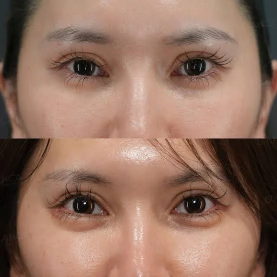
Reviewing unedited clinical canthoplasty before and after patient profiles demonstrates how repositioning the lateral canthal tendon restores eyelid contour and eye shape. The post-operative outcome reveals a lifted lower eyelid that rests against the lower border of the iris, eliminating scleral show and replacing a tired, rounded look with an elegant, stable almond eye contour.
Cost Determinants: Understanding Canthoplasty Pricing Variables
When evaluating lateral canthal surgery, prospective patients should note that pricing varies depending on individual anatomical complexity rather than a single flat fee. The overall canthoplasty cost is influenced by several clinical and logistical variables:
- Primary Aesthetic vs. Revisional Reconstructive Surgery: An uncomplicated aesthetic almond eye procedure involves different operative time and planning than a complex secondary reconstruction repairing lower lid retraction or scarring from a previous blepharoplasty.
- Combination Procedures: Canthoplasty is frequently performed alongside lower blepharoplasty (fat repositioning), midface lift, or endoscopic brow lift, altering overall theater time and surgical scope.
- Anesthesia & Surgical Setting: Procedures performed under local anesthesia with mild oral sedation in an outpatient suite involve different facility costs compared to twilight IV sedation or general anesthesia in an accredited hospital operating room.
- Surgeon's Specialization: Board-certified oculoplastic surgeons and facial plastic surgeons with dedicated ophthalmic training possess specialized micro-dissection skills to preserve corneal integrity, reflected in procedural fees.
- Comprehensive Logistics: All-inclusive medical tourism packages in Turkey incorporating VIP airport transfers, hotel accommodations, hospital stays, prescription eye drops, and follow-up examinations shape transparent pricing tiers.
Because periorbital structures and bone support are unique to each individual, exact quotations are established following a personalized clinical and photographic evaluation. The matrix below outlines relative regional price structures for 2026:
Healthcare Destination / Region | Cost Structure & Price Variability | Relative Price Level | Typical Package Framework |
United States (USA) | High variability by surgeon & city | Very High | Surgery Only (Facility, Drops & Anesthesia Extra) |
United Kingdom (UK) | Variable by private clinic & complexity | High | Surgery Only (Consultations & Checks Invoiced Separately) |
Germany / Switzerland | Variable by medical fee scale (GOÄ) | High | Surgery Only (Post-Op Drops & Hospitalization Extra) |
Stamboul (Turkey) | Dynamic Custom Package | High Value / Accessible | Comprehensive (Hospital, Hotel, VIP Transfers & Care) |
Frequently Asked Questions (FAQ)
Will my eyes look artificially slanted or pulled right after canthoplasty?
Yes, for the first two to four weeks, your eyes will appear slightly overcorrected and tightly slanted upward. Oculoplastic surgeons intentionally anchor the tendon slightly higher than the final desired position because tissues naturally relax downward by one to two millimeters as post-operative swelling resolves and internal scars mature. By weeks six to eight, the eyes soften into a natural, beautiful almond contour.
What is the primary difference between a canthoplasty and a blepharoplasty?
A lower blepharoplasty addresses excess skin and herniated fat pockets (under-eye bags) below the eye, but does not alter the structural position of the lateral corner. A canthoplasty cuts, tightens, and repositions the actual lateral canthal tendon to change the angle of the eye opening and elevate the lower lid margin. Both procedures are frequently combined to achieve comprehensive periorbital rejuvenation.
When can I safely wear contact lenses after canthoplasty surgery?
You must avoid wearing contact lenses for at least 3 to 4 weeks after surgery. Inserting and removing contact lenses pulls on the lower eyelid and can tear the delicate internal tarsal sutures away from the orbital bone before strong biological healing occurs. You should wear eyeglasses during this healing window.
Can canthoplasty correct a round eye shape caused by a previous bad surgery?
Yes. Post-blepharoplasty eyelid retraction, where too much skin or muscle was removed during an earlier lower eyelid surgery, leaving the eye rounded with visible scleral show, is one of the most common reasons for revisional canthoplasty. The surgeon releases internal scar contractures and reconstructs the tarsal tendon, restoring the lower lid's natural position against the eyeball.
Why do canthoplasty prices vary between different patients?
Pricing varies because treatment is tailored to the individual eyelid anatomy. Factors such as whether the procedure is an aesthetic primary lift or a complex revision of scar contractures, whether it is combined with lower blepharoplasty or midface lifting, the type of anesthesia used, and the inclusion of international travel packages all directly determine procedural duration and total cost.
Über den Autor